
Abdomen Wikipedia, la enciclopedia libre
Breasts. Summary. Female anatomy includes the external genitals, or the vulva, and the internal reproductive organs, which include the ovaries and the uterus. One major difference between males.
Abdominal Anatomy Pictures Female / 1896 Antique Medical Anatomy Print
The female sex organs consist of both internal and external genitalia. Together they comprise the female reproductive system, supporting sexual and reproductive activities. The external genital organs, or vulva, are held by the female perineum. These are the mons pubis, labia majora and minora, clitoris, vestibule, vestibular bulb and glands.

Female Abdominal Anatomy TrialExhibits Inc.
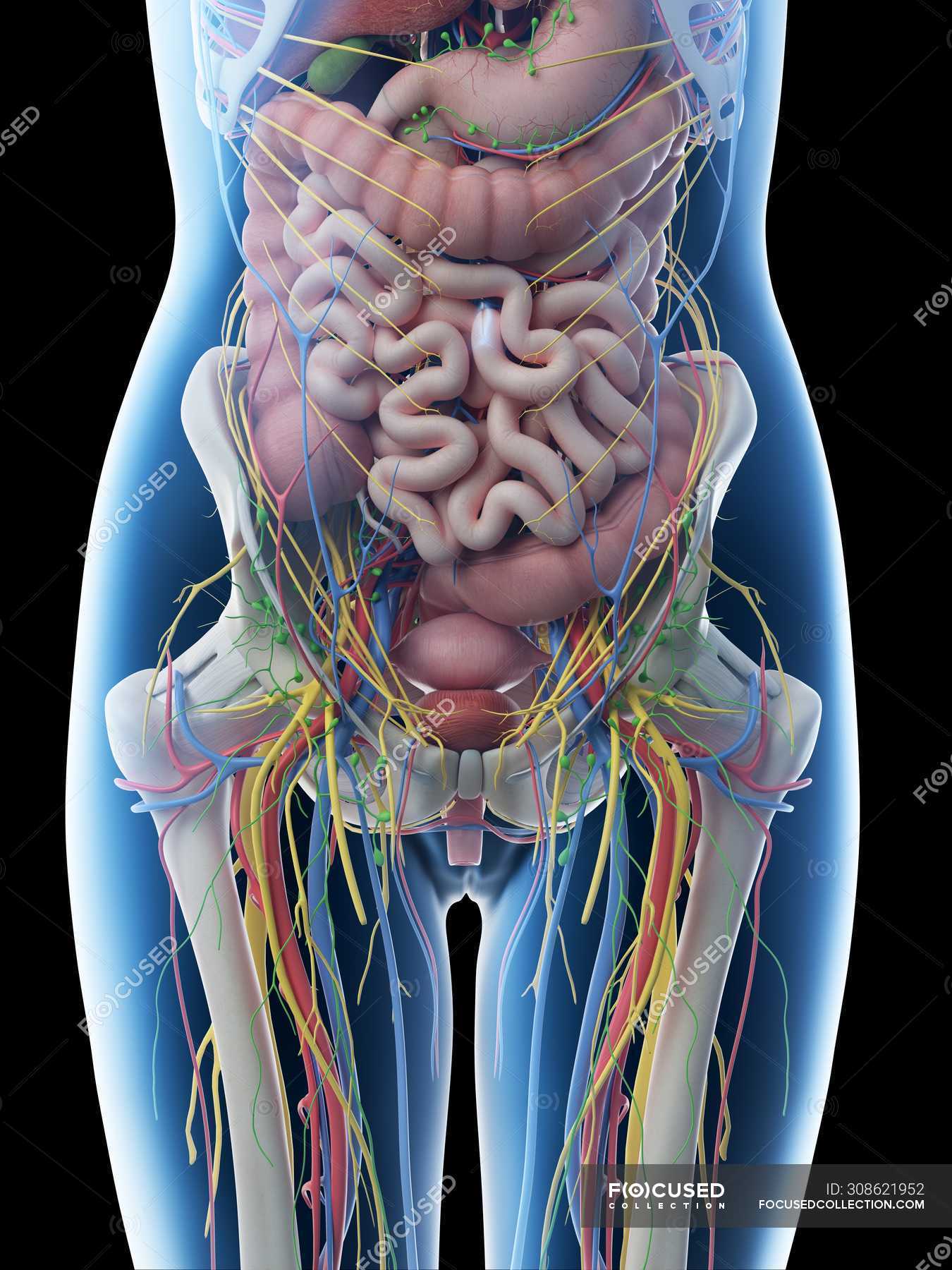
The abdomen and pelvic regions are continuous with each other, making up the distal part of the trunk. Bar the brain, heart and lungs, this region contains virtually all your body organs, including those involved in the digestive, endocrine, lymphatic, urinary and reproductive systems. So, it is crucial that you cover this section thoroughly.

lower abdomen anatomy diagram
Anatomy. Location. Function. Conditions. Tests. The uterus, also known as the womb, is a hollow, muscular organ located in the pelvis between the bladder and rectum of individuals who are assigned female at birth. This pear-shaped organ plays a role in menstruation, pregnancy, and childbirth. The lining of the uterus ( endometrium ) is the.
Abdominal Anatomy Pictures Female Abdominal Viscera Posterior Human
The bladder, also known as the urinary bladder, is an expandable, muscular sac that stores urine. When signaled, the bladder releases urine into the urethra, a tube that carries it out of the body.
Female Abdominal Anatomy, Artwork Photograph by Peter Gardiner
The abdominal muscles assist in the process of respiration, protect the inner organs, provide postural support, and serve to flex, extend, and rotate the trunk of the body. [4] The four main abdominal muscle groups, from innermost to outermost, can be remembered by the mnemonic TIRE: Transversus abdominis, internal oblique, rectus abdominis.
Anatomy Of The Female Abdomen And Pelvis, Cut away View Healthiack
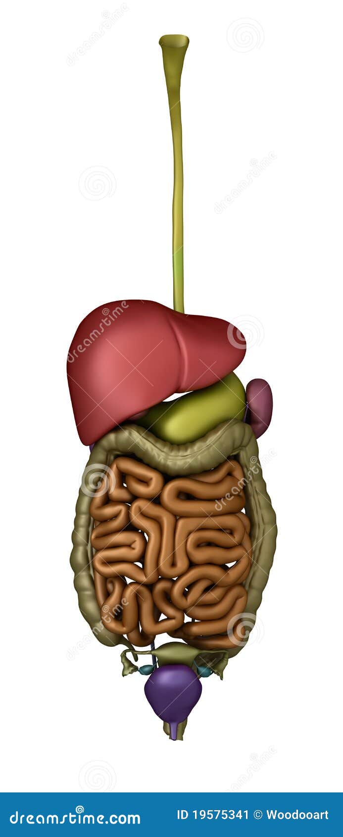
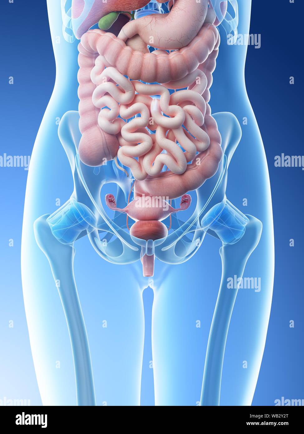
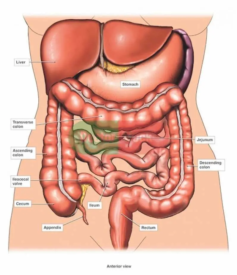
The abdomen contains organs involved in the gastrointestinal tract, including the oesophagus, stomach, small intestine, cecum, appendix, colon, rectum and the anal canal. The gastrointestinal tract is an organ system that enables us to ingest food, digest it, absorb it, and then expel the remaining waste as faeces.
Human Abdomen Anatomy Female Female Colon With Abdominal Organs
Peritoneum. Your peritoneum is a membrane that lines the inside of your abdomen and pelvis (parietal layer). It also covers many of your organs inside (visceral layer). The space in between these layers is called your peritoneal cavity. Folds of tissue form double layers, including your omentum, which hangs down the front of your abdomen, and.
/images/chapter/lymphatics-of-abdomen-and-pelvis/Lymphatics_of_abdomen_and_pelvis_2.png)
Abdominal Anatomy Organs Anatomy Of The Female Abdomen And Pelvis
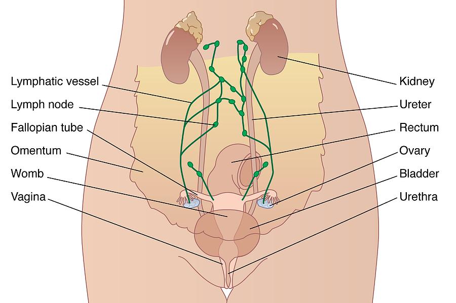
Uterus. Also called the womb, the uterus is a hollow, pear-shaped organ located in a woman's lower abdomen, between the bladder and the rectum. Ovaries. Two female reproductive organs located in the pelvis. Fallopian tubes. Carry eggs from the ovaries to the uterus. Cervix. The lower, narrow part of the uterus (womb) located between the bladder.

Major Organs In The Abdominal Cavity Elegant Of Human Abdominal Cavity
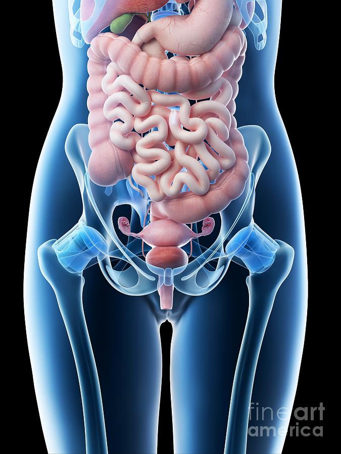
The pelvic cavity is a bowl-like structure that sits below the abdominal cavity. The true pelvis, or lesser pelvis, lies below the pelvic brim (Figure 1). This landmark begins at the level of the sacral promontory posteriorly and the pubic symphysis anteriorly. The space below contains the bladder, rectum, and part of the descending colon. In females, the pelvis also houses the uterus.
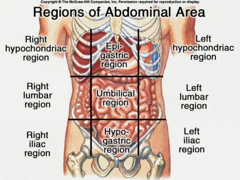
Female Abdomen Anatomy Quadrants / Abdominal Surface Anatomy Radiology
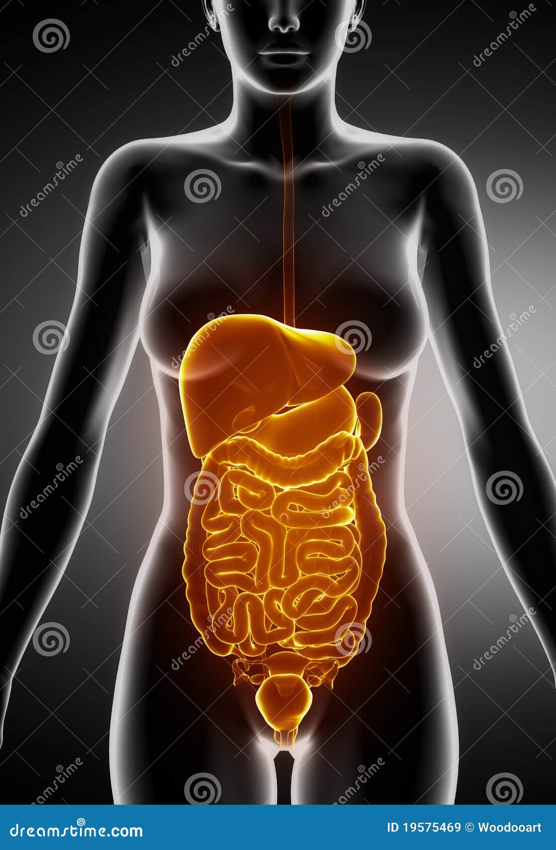
The abdomen contains many vital organs: the stomach, the small intestine (jejunum and ileum), the large intestine (colon), the liver, the spleen, the gallbladder, the pancreas, the uterus, the fallopian tubes, the ovaries, the kidneys, the ureters, the bladder, and many blood vessels (arteries and veins). Updated by: Debra G. Wechter, MD, FACS.
Female Anatomy Abdomen Images carfare.me 20192020
Summary. The five vital organs in the human body are the brain, heart, lungs, kidneys, and liver. Other organs include the gallbladder, pancreas, and stomach. Organ systems, such as the nervous.
Anatomy Of The Female Abdomen And Pelvis, Cut away View Healthiack
Anatomy of Female Pelvic Area. Click Image to Enlarge. Endometrium. The lining of the uterus. Uterus. Also called the womb, the uterus is a hollow, pear-shaped organ located in a woman's lower abdomen, between the bladder and the rectum. Ovaries. Two female reproductive organs located in the pelvis.

Human Anatomy Female, Human Anatomy Picture, Human Anatomy Chart, Human
The pelvis contains a large number of organs, bones, muscles, and ligaments, so many conditions can affect the entire pelvis or parts within it. Some conditions that can affect the female pelvis.
Female abdominal anatomy and internal organs, computer illustration
These internal structures of female anatomy include the: Vagina: The vagina is a muscular canal that connects the cervix and the uterus. It leads to the outside of the body. Parts of the vagina are made of collagen and elastin, which help it expand during sexual stimulation and childbirth. Cervix: The cervix is the lower part of the uterus that.
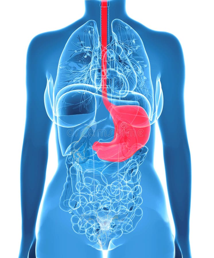
Female Abdomen Organs With Highlighted Stomach Stock Illustration
The female reproductive system is made up of external and internal organs. The external organs lie in an area called the vulva, and they include the labia, the clitoris, and the vaginal opening. The internal reproductive organs can be found within the pelvic cavity, and they include the ovaries, which produce the female sex cells, called oocytes, as well as sex hormones estrogen and.
